

Description
Date: Saturday 20.02.2027
Location: Panorama Hotel
Analipseos 26 Panorama,
Thessaloniki, PC 55236, Greece
Participation fee: €500
Maximum number of participants: 10
Scientific Director: Panagiotis Ermeidis, Rheumatologist
500,00 €
Ιnformational Seminar
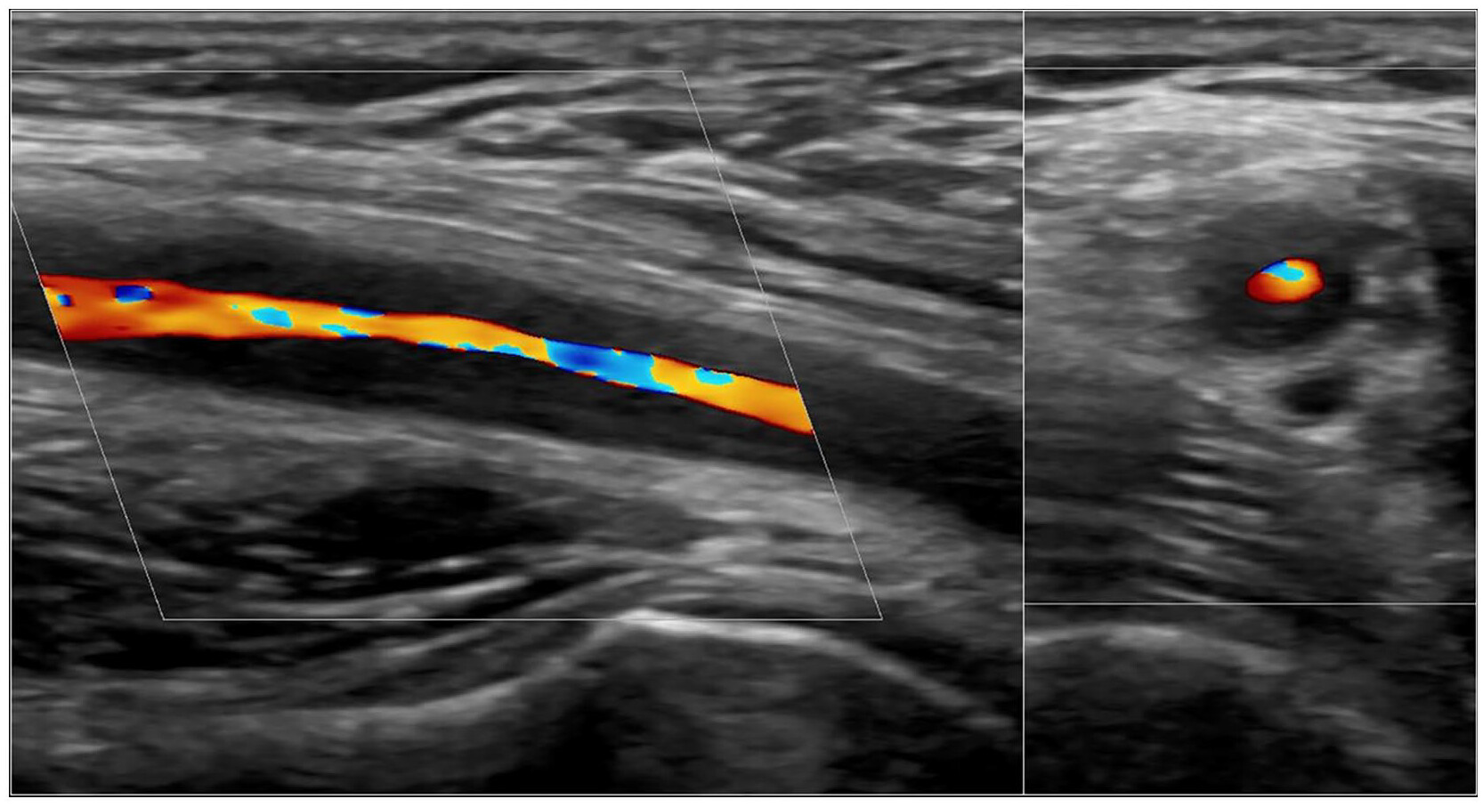
Ultrasound has evolved into an important tool for the early diagnosis and diagnostic assessment of Giant Cell Arteritis (GCA). Ultrasound examination of the temporal and axillary arteries is recommended by the European Alliance of Associations for Rheumatology (EULAR) as the first-line imaging modality in patients with suspected GCA, particularly in those presenting with cranial symptoms. Moreover, ultrasound findings are now incorporated into the classification criteria for the disease.
This one day seminar combines focused theoretical presentations with extensive hands-on sessions, allowing each participant to enhance their knowledge of ultrasound examination techniques, discuss clinical cases, and gain greater confidence in recognizing and interpreting ultrasonographic findings.
Why attend this seminar?
During the seminar, you will have the opportunity to:
Recognize the key ultrasonographic findings of GCA
Become familiar with the ultrasound assessment of the temporal and axillary arteries
Learn about internationally established ultrasound examination protocols
Correlate ultrasound findings with the clinical presentation
Understand the role of musculoskeletal ultrasound in Polymyalgia Rheumatica
Recognize findings that may contribute to the differential diagnosis
Practice hands-on, with expert guidance and dedicated time for individual practice
10 in stock
Date: Saturday 20.02.2027
Location: Panorama Hotel
Analipseos 26 Panorama,
Thessaloniki, PC 55236, Greece
Participation fee: €500
Maximum number of participants: 10
Scientific Director: Panagiotis Ermeidis, Rheumatologist
09:00 – 09:30 THEORETICAL SESSION I
PMR & GCA: An Introduction
Overview and clinical features of PMR and GCA
The role of ultrasound in the diagnosis and assessment of PMR and GCA
Ultrasound settings and optimisation for vascular examination
09:30 – 10:00 THEORETICAL SESSION II
Ultrasound in PMR: Shoulders, Hips & Differential Diagnosis
Ultrasound examination of the shoulder and hip regions
Typical sonographic findings in PMR
Ultrasound in the differential diagnosis of PMR
10:00 – 11:30 HANDS-ON SESSION I
PMR in Practice: Basic Sonoanatomy & Ultrasound Examination
Identification of key anatomical structures
Systematic ultrasound examination of the shoulder and hip
Recognition of characteristic sonographic findings
11:30 – 11:45 Coffee Break
11:45 – 12:15 THEORETICAL SESSION III
Ultrasound in GCA: Temporal & Cranial Arteries
Ultrasound examination of the temporal arteries
Examination of other cranial arteries
Normal and pathological sonographic findings in GCA
12:15 – 13:45 HANDS-ON SESSION II
GCA in Practice: Temporal & Cranial Artery Ultrasound
Practical examination technique
Systematic scanning approach
Identification of characteristic vascular findings
13:45 – 14:15 Lunch Break
14:15 – 14:45 THEORETICAL SESSION IV
Beyond the Temporal Arteries: Extracranial Vessel Ultrasound
Ultrasound examination of extracranial arteries in GCA
Vascular ultrasound in Takayasu Arteritis
Key sonographic findings and practical considerations
14:45 – 16:15 HANDS-ON SESSION III
Vascular Ultrasound in Practice
Hands-on examination of extracranial arteries
Practical scanning protocols
Recognition and interpretation of pathological vascular findings
16:15 – 16:30 FINAL QUIZ
Test Your Skills
Interactive quiz and review of the key learning points.
16:30 COURSE CONCLUSION
Summary, Discussion & Closing